RealNow
A one-stop AI assisted diagnostic platform for acute stroke
It is an imaging processing software that quickly obtains the imaging indicators required for stroke diagnosis and treatment. It can quickly provide accurate imaging analysis and quantitative data results for clinical decision-making on acute reperfusion treatment of stroke, assisting clinical rapid development of the best treatment plan.
According to the "Third National Sampling Survey Report on the Review of Causes of Death", cerebrovascular disease has now risen to the top cause of death in the country, with stroke being the disease with the highest disability rate of a single disease. One person in China experiences stroke every 12 seconds and one person dies from stroke every 21 seconds.
The current situation of stroke diagnosis and treatment in China: Traditional imaging processing takes a long time, lacks the quantitative imaging indicators required for ultra window thrombolysis and thrombectomy, relies on subjective experience, and is prone to misdiagnosis and misdiagnosis, resulting in high mortality and disability rates for stroke patients.
Product solutions
RealNow is a one-stop AI assisted diagnostic platform for acute stroke, which is an image processing software that quickly obtains the imaging indicators required for stroke diagnosis and treatment. It can provide accurate image analysis and quantitative data results for clinical decision-making on acute stroke reperfusion treatment, and assist in quickly developing the best treatment plan.
Comprehensive: Covering the analysis of imaging data required for the acute stage of stroke, providing a one-stop solution to the treatment decision-making needs of the acute stage, and assisting in clinical rational design of treatment plans.
Precision: Through multicenter clinical research, it has been shown that the consistency between AI assisted diagnostic data results and the diagnostic results of doctors with 20 years of clinical experience is over 95%.
Fast: Complete imaging analysis within 3 minutes, quickly provide auxiliary diagnostic results, make quick decisions, treat quickly, and save valuable acute treatment time.
Objective: AI algorithm intelligent analysis eliminates subjectivity in the diagnosis process of different doctors, and outputs objective and consistent quantitative indicators.
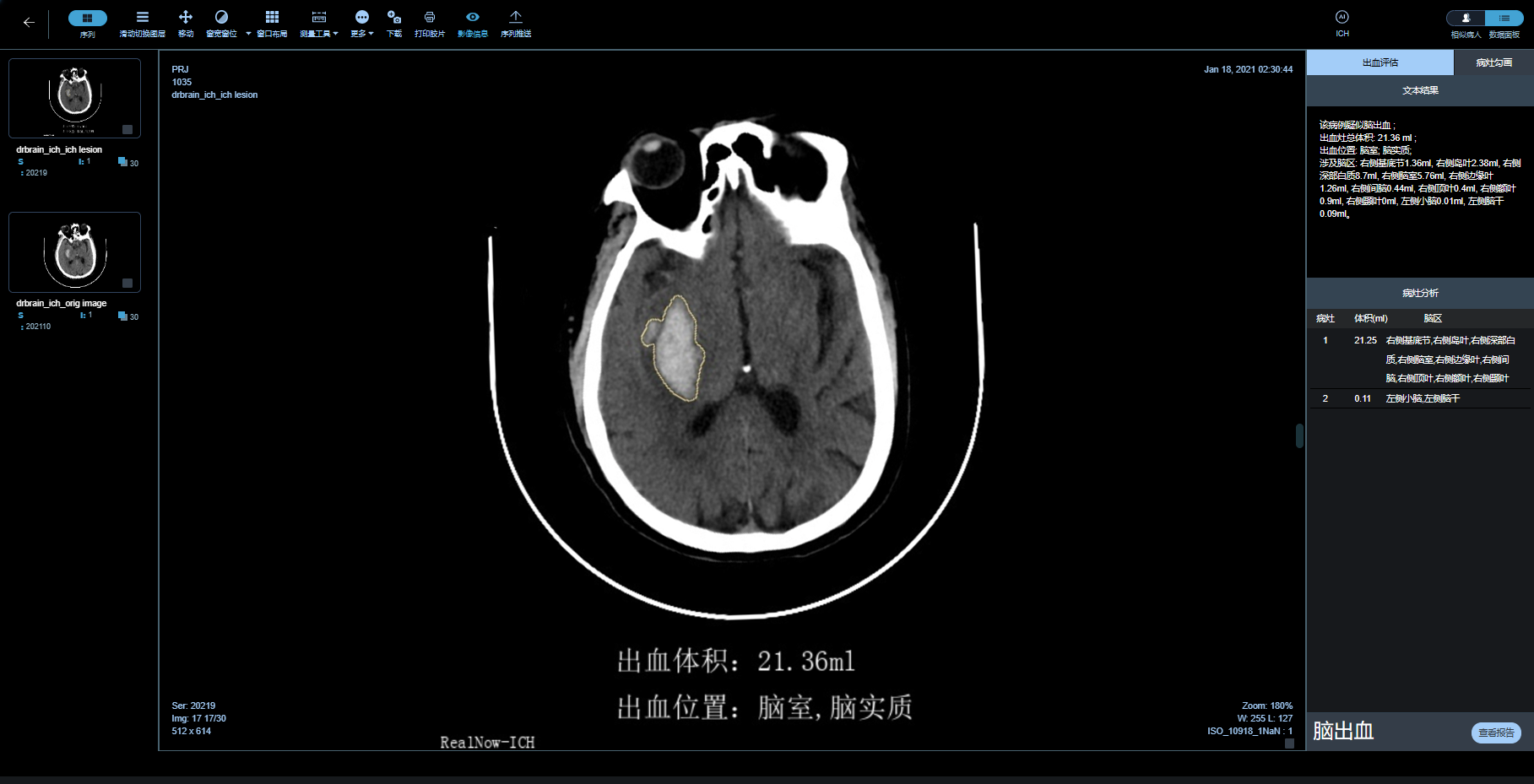
Intelligent analysis of CT plain scan (NCCT) images, rapid and intelligent analysis of intracranial hemorrhage, identification of hematoma location, calculation of hematoma volume, and assistance to clinical doctors in making treatment decisions quickly.
Assisted decision-making of treatment plans for hemorrhagic stroke - Intelligent identification and quantitative evaluation of bleeding foci
Analysis of bleeding site and type
Automatically complete precise delineation of hematoma boundaries
Automatically provide accurate hematoma volume values
Automatic analysis of midline deviation in the brain
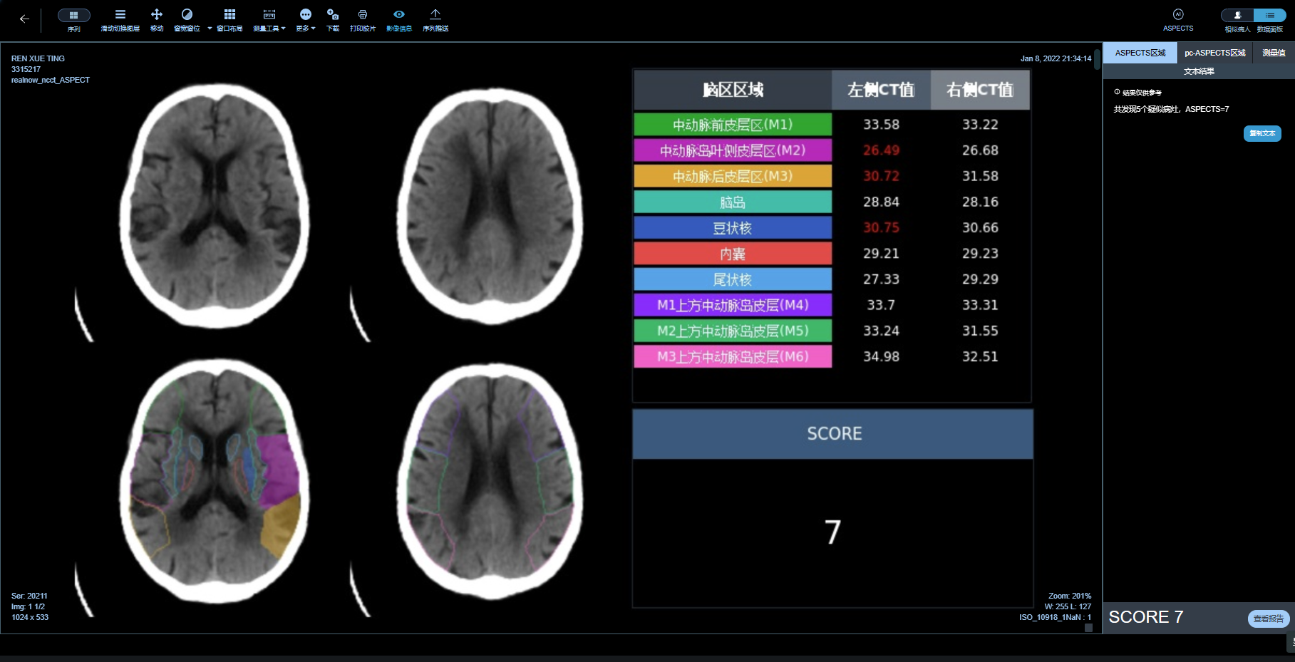
Identification of new infarct lesions and ASPECTS score for lesion severity. AI automatically and quickly provides ASPECTS scores for the 10 functional areas of the anterior circulation and the 8 functional areas of the posterior circulation, solving the problems of long time and poor consistency when doctors manually select brain regions for scoring. This provides a key auxiliary decision-making basis for clinical doctors to formulate treatment plans for ischemic stroke。
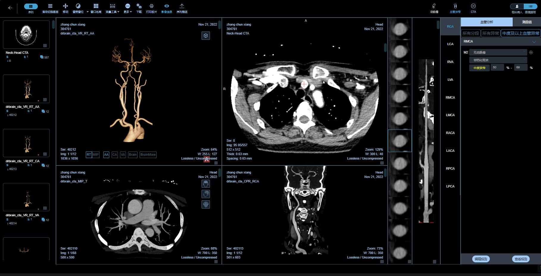
Through rapid and automatic analysis of CTA images, key auxiliary basis is provided for clinical intervention treatments such as mechanical thrombectomy.
Assisted decision-making of mechanical thrombectomy treatment plan within 6 hours - vascular stenosis assessment and surgical path planning
Intelligent analysis of CTA images through AI:
Automatic head and neck vascular reconstruction
Identify the location of vascular stenosis
Assess the degree of vascular stenosis
Automatic vascular segmentation labeling
Identification of atherosclerotic plaque
Fully automatic aneurysm detection
Cerebrovascular centerline editing
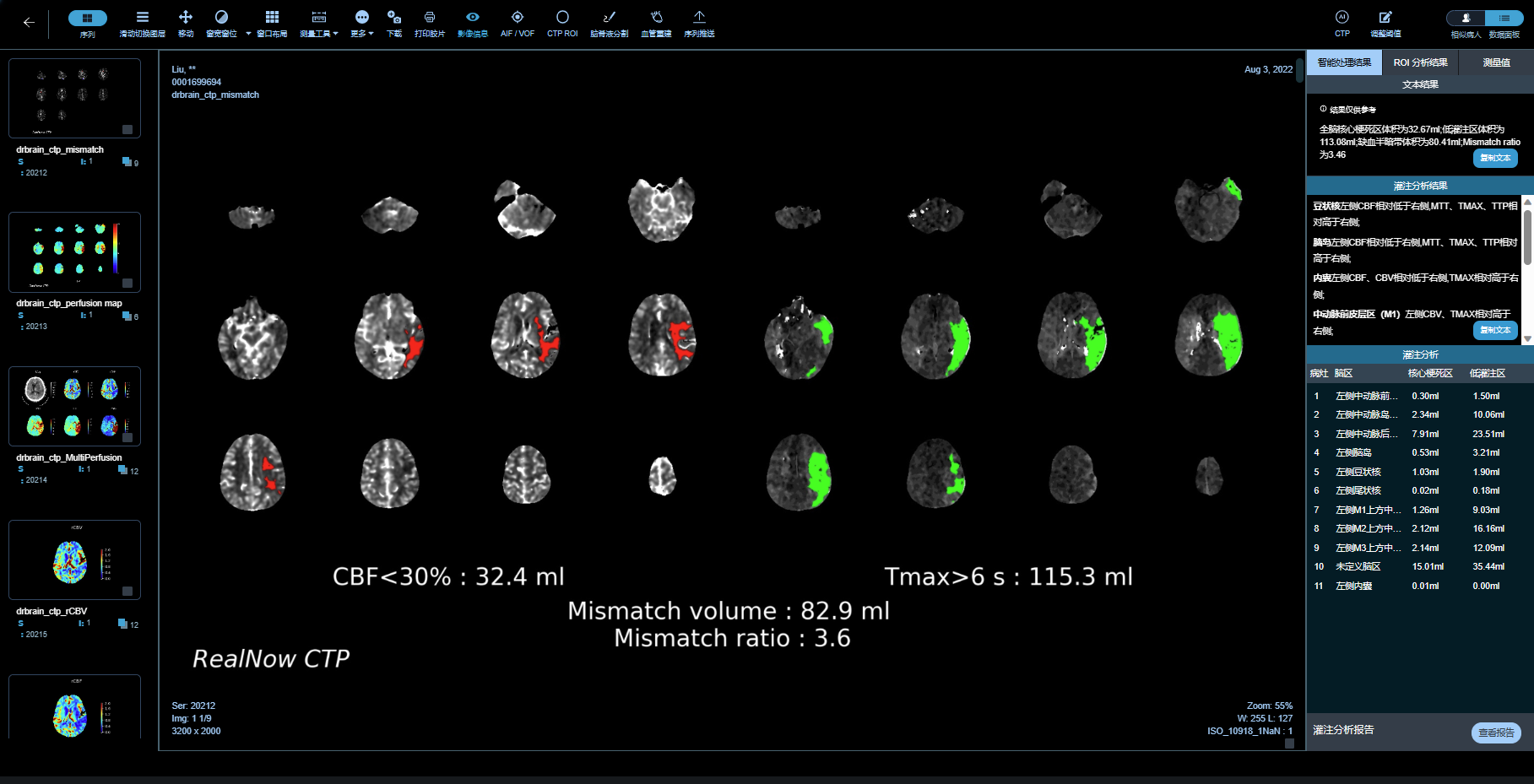
6 hour external ultrasound window thrombectomy treatment and assisted decision-making for stroke treatment plan after awakening - intelligent fully automatic mismatch value evaluation
Support multimodal automatic organization window perfusion analysis and data display
Automatic display of core infarct area, ischemic penumbra, and automatic mismatch value calculation provide objective and quantitative surgical indications for clinical ultrasound thrombectomy and stroke after awakening.
Provide analysis of multiple CBF threshold core infarct areas and multiple Tmax threshold ischemic penumbra, as well as comparison of ischemic penumbra and core infarct areas under different thresholds, to assist clinical assessment of patients' condition from multiple perspectives and adapt to scientific research needs.
Rapid three-dimensional reconstruction of cerebral blood vessels based on CTP imaging data meets the clinical needs of cerebrovascular intervention doctors in evaluating the condition of cerebral blood vessels from multiple perspectives.
Fully automatic cerebrospinal fluid segmentation and head movement quantification correction, providing more accurate cerebral perfusion quantification data.
More widespread CTP imaging and more accurate DWI/PWI imaging for full coverage, enabling multimodal intelligent image analysis.